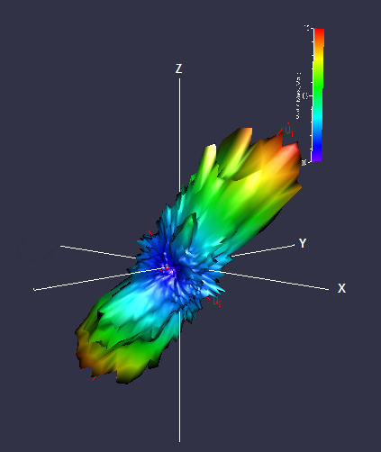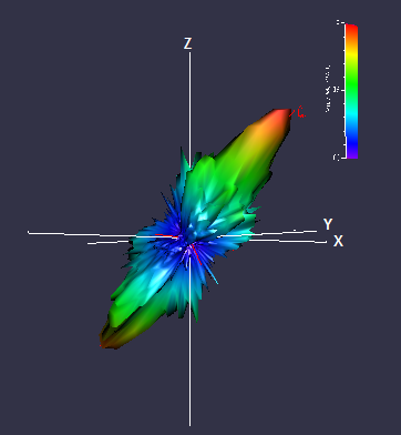

X-ray computed tomography is a powerful and non-destructive technique for studying the three-dimensional (3D) architecture of trabecular bone. The images show 3D rose diagrams obtained from analysing CT scans of femoral necks, using Quant 3D and a Star Volume Distribution (SVD) method. These diagrams show the average shape of trabeculae in femoral necks; top: plate-like and bottom: rod-like. Also shown on each diagram are the main axes of symmetry as red lines. The diagrams are coloured to allow relative length comparison.
| Dr. Athina Markaki |
Department of Engineering |
| Dr. Paul Mayhew |
Department of Medicine |
| Eve Mullen |
|
| Dr. Ken Poole |
Department of Medicine |
| Dimitris Tsarouchas |
Department of Engineering |

 X-ray computed tomography is a powerful and non-destructive technique for studying the three-dimensional (3D) architecture of trabecular bone. The images show 3D rose diagrams obtained from analysing CT scans of femoral necks, using Quant 3D and a Star Volume Distribution (SVD) method. These diagrams show the average shape of trabeculae in femoral necks; top: plate-like and bottom: rod-like. Also shown on each diagram are the main axes of symmetry as red lines. The diagrams are coloured to allow relative length comparison.