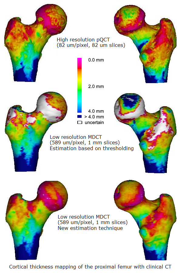
The distribution of cortical bone (the denser outer layer of bone) in the hip is believed to be critical in determining fracture resistance. Generally cortical bone around the femoral neck gets thinner with age, and can become as thin as an egg-shell (0.3 mm) by the eighth decade in critical areas. Current clinical practice is to measure overall hip bone mineral density (called a 'DXA' scan) which is not very sensitive to cortical thickness. Potentially, multi-detector computed tomography (MDCT) can be used to examine cortical thickness and 3-D structure directly. However, existing CT technology has a limited resolution, which is not good enough to be able to distinguish the bone cortex, particularly in clinically relevant thin cases.

A collaboration between the Engineering Department and the Department of Medicine has led to the development of an algorithm which can accurately measure cortical thickness from clinical MDCT data and display this as a colour map over the bone surface. This has been demonstrated to be much more accurate than existing techniques for cortical assessment.
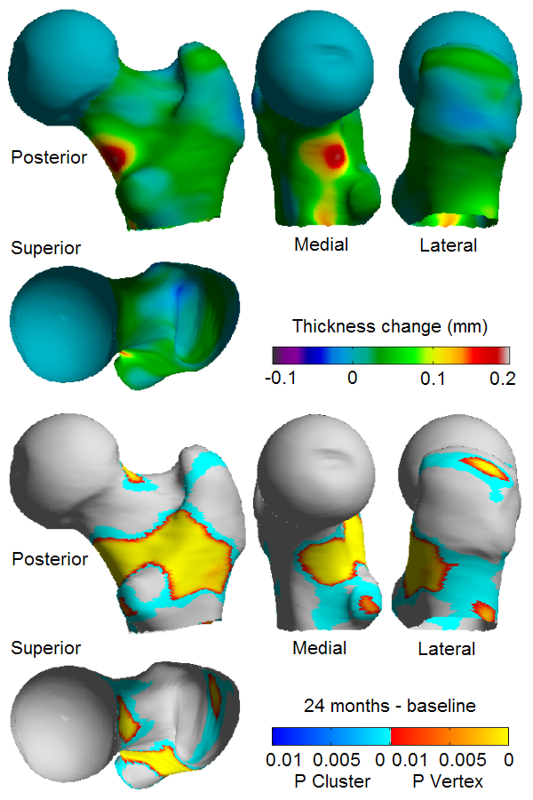
This new tool opens up many research possibilities. For instance a recent study on the treatment of bone with Teriparatide has already been able to demonstrate not only statistically significant bone growth in the 0.05 - 0.1 mm range, but also exactly where the bone growth is occuring. There are fascinating parallels between these areas and the stress distribution and muscle insertion points on the femur.
| Dr .Andrew Gee |
Department of Engineering |
| Dr. Paul Mayhew |
Department of Medicine |
| Dr. Ken Poole |
Department of Medicine |
| Dr. Graham Treece |
Department of Engineering |
 A collaboration between the Engineering Department and the Department of Medicine has led to the development of an algorithm which can accurately measure cortical thickness from clinical MDCT data and display this as a colour map over the bone surface. This has been demonstrated to be much more accurate than existing techniques for cortical assessment.
This new tool opens up many research possibilities. For instance a recent study on the treatment of bone with Teriparatide has already been able to demonstrate not only statistically significant bone growth in the 0.05 - 0.1 mm range, but also exactly where the bone growth is occuring. There are fascinating parallels between these areas and the stress distribution and muscle insertion points on the femur.