The term elastography refers to a range of techniques that attempt to measure tissue stiffness, which has long been recognised as a useful indicator of disease. This is done by deforming the tissue: stiff lesions deform less than their surroundings and are often readily apparent in the resulting elastograms. In freehand quasistatic ultrasonic strain imaging, the deformation is applied by manually pressing on the ultrasound probe and then measured by analysing the ultrasound data. Although this is a qualitative technique that is fundamentally incapable of measuring absolute tissue stiffness, it is appealing in that it requires only conventional ultrasound scanning hardware and a number of manufacturers have recently started to offer this sort of elastography on their flagship machines.
Researchers at the Engineering Department have developed a novel elastography system that produces particularly high resolution, stable images, with regions of poor stiffness measurement hidden with an overlaid colour mask. The colour mask reduces the potential to misinterpret noise as meaningful stiffness information, and also helps to differentiate cystic and solid lesions. This tool is being investigated for scanning the breast, liver, musculoskeletal system, pelvis, placenta and neck.

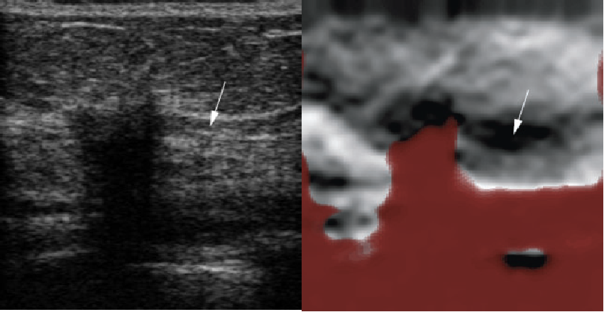
The left hand B-mode image shows an invasive breast carcinoma with ductal carcinoma in situ (DCIS). The adjacent intra-ductal extension (arrow) is more obvious in the strain image: lighter shades indicate soft whereas darker shades indicate stiff areas. The red wash in the strain image masks noise caused by loss of signal under the lesion and towards the bottom of the frame.

This is another invasive breast carcinoma, with the left hand B-mode image showing a typical irregular breast mass. The strain image shows a corresponding stiff irregular lesion, but with an unexpected area of red masking in the centre. This masking turned out from a core biopsy to correspond to an area of necrosis.
| Dr. Lol Berman |
Department of Radiology |
| Dr. Trish Chudleigh |
Lead Sonography, Rosie Maternity Hospital |
| Dr. Robin Crawford |
Consultant Gynaecologist and Oncologist, NHS |
| Dr .Andrew Gee |
Department of Engineering |
| Dr. Melanie Hopper |
Consultant Radiologist, NHS |
| Prof. David Lomas |
Department of Radiology |
| Prof. Richard Prager |
Department of Engineering |
| Dr. Evis Sala |
Department of Radiology |
| Dr. Ruchi Sinnatamby |
Consultant Radiologist, NHS |
| Kathryn Taylor |
Advanced Practitioner Radiographer, NHS |
| Dr. Graham Treece |
Department of Engineering |
 The left hand B-mode image shows an invasive breast carcinoma with ductal carcinoma in situ (DCIS). The adjacent intra-ductal extension (arrow) is more obvious in the strain image: lighter shades indicate soft whereas darker shades indicate stiff areas. The red wash in the strain image masks noise caused by loss of signal under the lesion and towards the bottom of the frame.
 This is another invasive breast carcinoma, with the left hand B-mode image showing a typical irregular breast mass. The strain image shows a corresponding stiff irregular lesion, but with an unexpected area of red masking in the centre. This masking turned out from a core biopsy to correspond to an area of necrosis.
 The left hand B-mode image shows an invasive breast carcinoma with ductal carcinoma in situ (DCIS). The adjacent intra-ductal extension (arrow) is more obvious in the strain image: lighter shades indicate soft whereas darker shades indicate stiff areas. The red wash in the strain image masks noise caused by loss of signal under the lesion and towards the bottom of the frame.
 This is another invasive breast carcinoma, with the left hand B-mode image showing a typical irregular breast mass. The strain image shows a corresponding stiff irregular lesion, but with an unexpected area of red masking in the centre. This masking turned out from a core biopsy to correspond to an area of necrosis.